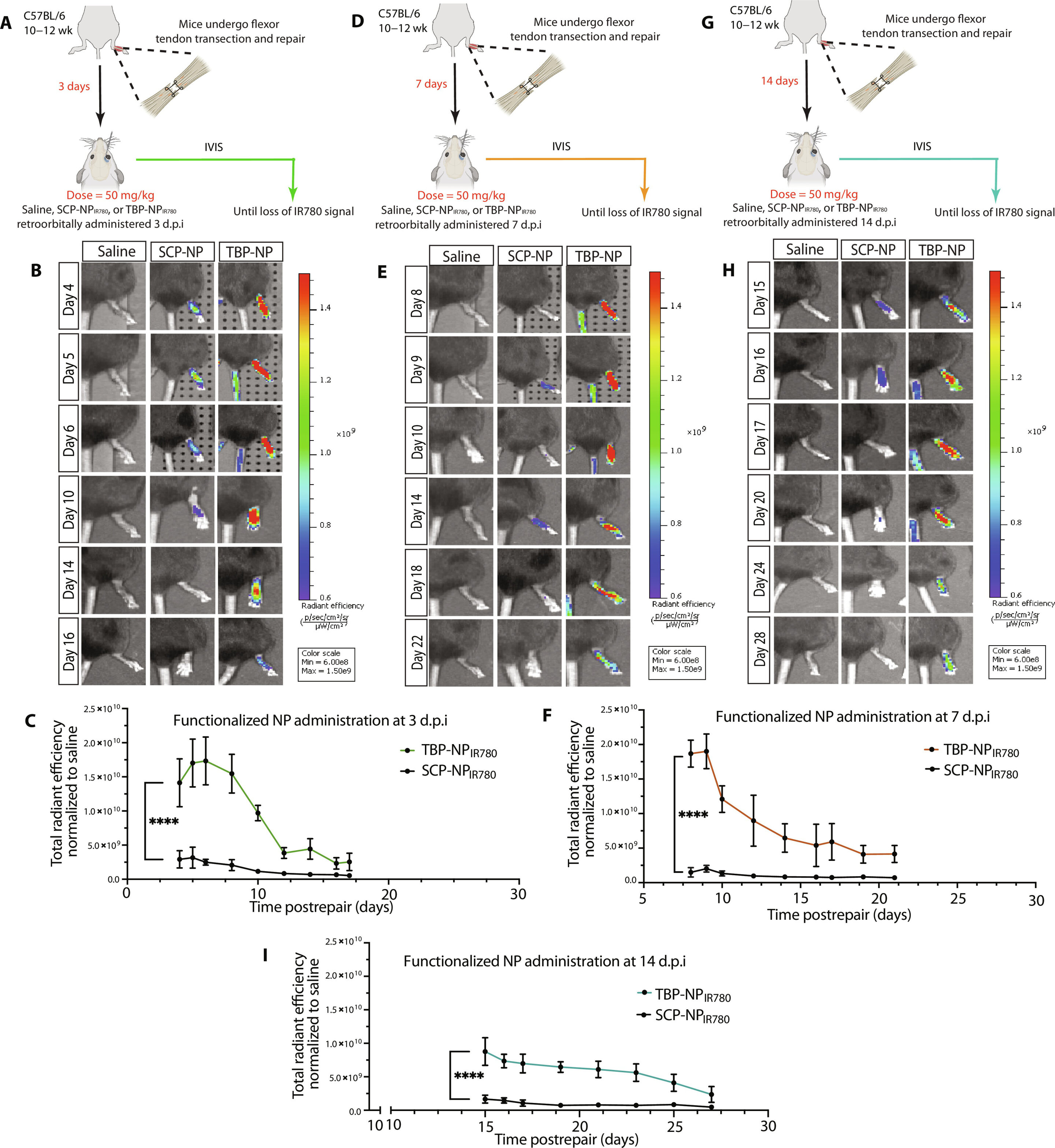
Use of a TBP-functionalized NP delivery system to target injured tendons. (A) Schematic representation of the treatment schedule for targeted studies 3 days post-injury. (B and C) Representative images and graphs of live animals showing the biodistribution of NPs after treatment on day 3. (D) Schematic representation of the treatment schedule for targeted studies 7 days (E and F) Representative images and graphs of live animals showing the biodistribution of NPs after treatment on day 7. (G) Schematic representation of the treatment schedule for targeted studies 14 days (H and I) Representative images and graphs of live animals showing the biodistribution of NPs after treatment on day 14. Image credit: Scientific advances (2024). DOI: 10.1126/sciadv.adn2332
× close
Use of a TBP-functionalized NP delivery system to target injured tendons. (A) Schematic representation of the treatment schedule for targeted studies 3 days post-injury. (B and C) Representative images and graphs of live animals showing the biodistribution of NPs after treatment on day 3. (D) Schematic representation of the treatment schedule for targeted studies 7 days (E and F) Representative images and graphs of live animals showing the biodistribution of NPs after treatment on day 7. (G) Schematic representation of the treatment schedule for targeted studies 14 days (H and I) Representative images and graphs of live animals showing the biodistribution of NPs after treatment on day 14. Image credit: Scientific advances (2024). DOI: 10.1126/sciadv.adn2332
Using nanoparticles to deliver drugs to a surgically repaired tendon is a promising new approach that reduces scar tissue formation and improves mechanical function. The researchers’ success in localizing drug therapy in the body at the cellular level proved to be a highly efficient delivery method that could be used to treat other injuries. Their study was published in Scientific advances.
Whether it’s a season-ending Achilles tendon tear suffered by professional football player Aaron Rodgers or a routine workplace accident, tendon injuries are common and can be life-changing. They require 300,000 surgeries each year and frequently result in lost work time and permanent physical disability.
Typically, traumatic tendon injuries are repaired surgically with sutures. However, optimal healing is often compromised by the tendon’s tendency to form scar tissue, which limits the tendon’s movement and function.
Researchers at the University of Rochester and the University of Oregon have combined their expertise in tendon cell biology and drug delivery systems to find a better method for therapies that can reduce scar tissue and promote healing.
“Despite the high number of these injuries and the often poor outcomes, there are very few effective medications to aid the tendon healing process,” said Alayna Loiselle, Ph.D., associate professor at the University of Rochester’s Center for Musculoskeletal Research.
“Systemic drug treatments administered orally or by injection have poor tendon adaptation; in some cases, less than 1% of a systemically administered drug reaches the healing tendon. Local administration of drugs directly to the tendon also has disadvantages, including potential tissue damage from the injection and poor control of drug concentrations at the injury site.”
“We want to move from suturing alone to incorporating therapeutics,” said Emmanuela Adjei-Sowah, a biomedical engineering doctoral student at the University of Rochester who has spent the past few years working with co-authors Loiselle, Danielle SW Benoit, Ph.D., and others to develop a nanoparticle delivery system to improve healing in tendon injuries. “Advances in multiomics and nanoparticle drug delivery are opening up new treatment options.”
The challenge for the researchers was to find out which substances could support tendon healing.
Molecular “map” of the healing process shows new therapeutic path
“The fundamental cellular and molecular mechanisms that drive tendon healing through scars are only just beginning to be clearly defined,” said Loiselle. “Our previous work used spatial transcriptome profiling to create a molecular map of the healing tendon. When we subsequently analyzed these data, we found that the area directly at the injury site had high Acp5 gene expression, which was both surprising and exciting.”
The Acp5 gene produces a protein called tartrate-resistant acid phosphatase (TRAP); both are known to occur in the recovery and rebuilding of injured bone. High TRAP activity within the healing tendon allowed researchers to use a peptide that binds to TRAP to deliver drugs directly to the healing tendon site.
“While Benoit’s lab has previously used TRAP-binding peptide nanoparticles (TBP-NPs) for targeted drug delivery in bone, the high TRAP activity in the healing tendon opened up a whole new line of research,” said Loiselle.
Before beginning trials of therapeutic agents, the team conducted a series of dosing and timing studies using a mouse model with complete transection and surgical repair of the flexor tendon to define the window of time during which their drug delivery system can most effectively reach the healing tendon.
“Defining an optimal treatment window is critical to the successful development of an innovative and effective drug delivery system that improves tendon healing by stimulating a regenerative rather than fibrotic healing cascade,” said Benoit, Lorry Lokey Department Chair and Professor in the Department of Bioengineering at the University of Oregon.
“By defining the optimal therapeutic window for this drug delivery system, we can also mitigate the unwanted side effects typically associated with the high doses or multiple doses required to achieve adequate tissue accumulation of the drug.”
As a therapeutic, the team chose niclosamide, which inhibits S100a4, a protein that Loiselle’s lab had already identified as contributing to scar formation. Previous work from Loiselle’s lab showed that genetically knocking out S100a4 improved mechanical and functional outcomes in this tendon healing model in mice.
S100a4 is associated with scar formation in numerous tissues, including the liver, heart, lung, and oral submucosa. Loiselle’s discovery that it impedes tendon healing provided a therapeutic target for this study, where they used their drug delivery system based on TRAP-binding peptide nanoparticles to specifically target the injured tendon.
For comparison, they administered niclosamide systemically. This slightly reduced the amount of S100a4 in the healing tendon but had no positive effect on the healing process. In contrast, administration of the same dose of niclosamide using the nanoparticle system resulted in a strong inhibition of S100a4 mRNA and protein levels in the healing tendon.
This targeted delivery method of the drug also had a significant positive impact on the tendon healing process. Delivery of niclosamide through TBP-NP improved both the recovery of functional range of motion and the mechanical integrity of the healing tendon over both short and long periods of time. Importantly, these sustained effects occurred after just a single treatment.
The researchers will continue their work to define the broad scope to which the system can be used for other tendon injuries and diseases, as well as other types of tissue injuries that lead to scarring.
“The beauty of this system is that it can be loaded with different types of drugs to target different molecular processes or pathways,” said Adjei-Sowah.
More information:
Emmanuela Adjei-Sowah et al., Development of a nanoparticle-based tendon-targeted drug delivery system for pharmacological modulation of tendon healing, Scientific advances (2024). DOI: 10.1126/sciadv.adn2332
Information about the magazine:
Scientific advances